

Key Takeaways
- Mammograms are the primary recommended screening tool for most women starting at age 40, per major clinical guidelines.
- Breast ultrasound is typically used as a diagnostic complement, not a standalone routine screening replacement.
- Dense breast tissue can limit mammogram sensitivity, making ultrasound a useful supplemental tool in those cases.
- Neither test is universally superior — clinicians choose based on individual risk, tissue type, and clinical findings.
- Always discuss your personal screening plan with a qualified healthcare provider.
Option A
Mammogram
The established first-line screening standard for breast health.
Best for: Routine screening in average-risk women, especially those 40 and older, and for detecting calcifications or early structural changes.
Option B
Breast Ultrasound
The targeted, radiation-free complement for specific diagnostic questions.
Best for: Distinguishing solid masses from fluid-filled cysts, evaluating dense breast tissue, and guiding biopsies or follow-up imaging.
If you are an average-risk woman seeking routine annual breast screening
Mammogram
Mammography is backed by decades of evidence for detecting early breast changes in asymptomatic women and remains the standard of care for routine screening.
If a mammogram has identified an area of concern requiring further evaluation
Breast Ultrasound
Ultrasound excels at characterizing whether a detected abnormality is a solid mass or a benign fluid-filled cyst, guiding next steps.
If you have been told you have dense breast tissue
Breast Ultrasound
Supplemental ultrasound can improve detection rates in dense breasts, where mammogram sensitivity is often reduced.
If you are pregnant or need to avoid ionizing radiation
Breast Ultrasound
Ultrasound uses sound waves and involves no radiation, making it the preferred imaging option during pregnancy or when radiation exposure is a concern.
How Each Technology Works
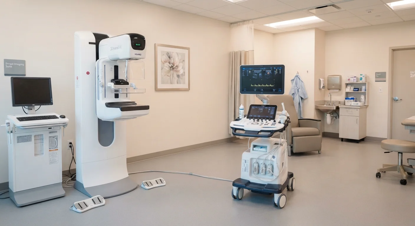
A mammogram is an X-ray of breast tissue. The breast is briefly compressed between two plates while low-dose ionizing radiation produces a detailed image highlighting differences in tissue density, structural changes, and tiny calcium deposits called microcalcifications. These calcifications can be early indicators of certain breast conditions and are often invisible to other modalities.
A breast ultrasound uses high-frequency sound waves emitted from a handheld probe (transducer) pressed against the skin. As sound waves bounce back from breast structures, a computer translates the echoes into real-time images. Because ultrasound does not use radiation, it is safe during pregnancy and can be repeated without cumulative radiation exposure.
Understanding how each tool functions helps clarify why clinicians reach for one over the other — and why they are often used in sequence rather than as substitutes. See how breast awareness tools work together for a broader picture of layered breast health monitoring.
| Criterion | Mammogram | Breast Ultrasound |
|---|---|---|
| Technology | Low-dose X-ray (ionizing radiation) | High-frequency sound waves (no radiation) |
| Primary use | Routine population screening | Diagnostic follow-up, dense tissue, symptoms |
| Detects calcifications | Yes — a key strength | Poorly — a significant limitation |
| Performance in dense tissue | Reduced sensitivity | Less affected by tissue density |
| Distinguishes cysts from solid masses | Limited | Yes — a core strength |
| Safe during pregnancy | Generally avoided; used only if clinically urgent | Yes — preferred in pregnancy |
| Typical session duration | 10–20 minutes | 15–30 minutes |
| Biopsy guidance | Stereotactic biopsy only | Routinely used for real-time guidance |
When Clinicians Choose One Over the Other
Mammography is the only imaging modality with robust, long-term evidence supporting its use as a population-level screening tool. Major organizations including the American College of Obstetricians and Gynecologists (ACOG) and the American Cancer Society have issued guidelines recommending regular mammograms for average-risk women, typically beginning at age 40 — though recommendations vary by organization and individual risk profile.
Breast ultrasound serves a different clinical role. It is most commonly ordered in the following situations:
- Diagnostic follow-up: When a mammogram flags an area of concern, ultrasound helps characterize it — determining whether the finding is a simple cyst (fluid-filled and generally benign) or a solid mass requiring further investigation.
- Dense breast tissue: Approximately 40% of women have dense breasts, according to the National Cancer Institute. Dense tissue appears white on mammograms, as do tumors, which can obscure findings. Ultrasound uses a different contrast mechanism that is not affected by tissue density in the same way.
- Symptomatic evaluation: If a woman or clinician notices a palpable lump, nipple discharge, or localized pain, ultrasound provides rapid, targeted assessment.
- Biopsy guidance: Ultrasound is routinely used in real time to guide needles during minimally invasive breast biopsies.
For women at elevated risk due to genetic factors, personal or family history, ultrasound may be recommended alongside mammography — and MRI may also enter the conversation. Your provider is best positioned to tailor that plan. Learn what each routine women's health screening actually measures to understand where breast imaging fits in your broader preventive care.
~40%
Women with dense breast tissue in the U.S.
According to the National Cancer Institute, dense breast tissue affects a substantial portion of women and can limit mammogram sensitivity.
87%
Approximate mammogram sensitivity in non-dense tissue
Sensitivity can drop significantly in women with extremely dense breasts, underscoring the clinical rationale for supplemental ultrasound.
Limitations, Risks, and What to Expect
Both modalities have known limitations that clinicians weigh when making recommendations.
Mammogram considerations: False positives can occur — findings that prompt additional imaging or biopsy but ultimately prove benign. This can cause anxiety and additional procedures. Conversely, false negatives (missed cancers) are also possible, particularly in dense tissue. The radiation dose per mammogram is low, but it is not zero.
Ultrasound considerations: Ultrasound is highly operator-dependent; image quality and interpretation vary with the skill and experience of the technician and radiologist. It is less sensitive to microcalcifications — an important early finding — making it a poor substitute for mammography as a primary screening tool for most women.
Neither test is perfectly sensitive or specific. A normal result does not guarantee the absence of disease, and an abnormal result does not confirm it. This is why ongoing, guideline-driven screening — not one-time testing — is the foundation of proactive breast health. Explore the full range of reproductive health screenings every adult woman should understand.
This article is for general informational and educational purposes only and does not constitute medical advice. Breast imaging decisions should always be made in consultation with a qualified healthcare provider who can account for your individual health history, risk factors, and clinical findings.