

How a Urine Dipstick Works
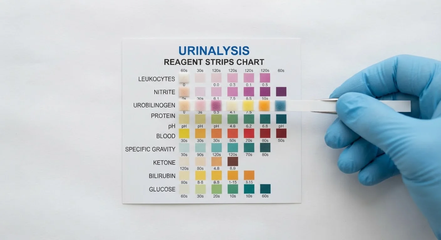
A urine dipstick is a narrow plastic strip carrying up to 10–12 small, chemically treated pads. Each pad is impregnated with reagents that react specifically to one substance — such as glucose, protein, or blood — present in a urine sample. When the strip is briefly dipped into fresh urine and then removed, each pad changes colour (or stays the same) within a defined time window, typically 30 to 120 seconds depending on the marker.
Reading the strip means holding it horizontally to prevent colours from running between pads, then comparing each pad against the colour scale printed on the container label — not a separate booklet — at the exact recommended time. Timing matters: reading a pad too early or too late can shift the apparent result.
Dipstick tests are qualitative or semi-quantitative. They indicate whether a substance is absent, trace, or present at a certain approximate level — they do not give precise numerical concentrations the way a laboratory analyser does. For that reason, any abnormal finding on a dipstick should be confirmed by a healthcare professional using laboratory urinalysis or follow-up testing.
What Each Pad on the Colour Chart Measures
Most multi-parameter dipsticks test for the following markers. Results are expressed as negative, trace, or graded levels (1+, 2+, 3+).
- pH (acidity/alkalinity): Normal urine ranges roughly from pH 4.5 to 8.0. Very alkaline urine can indicate certain kidney conditions or a urinary tract infection (UTI) caused by urea-splitting bacteria; very acidic urine may reflect diet or metabolic states.
- Specific gravity: Reflects urine concentration — how well the kidneys are diluting or concentrating urine. Low values may suggest excess fluid intake or impaired kidney concentration; high values may indicate dehydration.
- Protein (albumin): Healthy urine contains little or no protein. A trace or positive result can signal kidney stress, UTI, or conditions such as chronic kidney disease, though strenuous exercise and fever can also cause temporary elevations.
- Glucose: Glucose should not normally appear in urine. Its presence (glucosuria) may suggest blood glucose has exceeded the kidney's reabsorption threshold — a common finding in uncontrolled diabetes. See how blood glucose monitors complement this picture for context.
- Ketones: Detected when the body is breaking down fat for fuel, as in prolonged fasting, very low-carbohydrate diets, or uncontrolled type 1 diabetes (diabetic ketoacidosis).
- Blood (haematuria): Even trace blood in urine warrants follow-up — causes range from UTI and kidney stones to more serious conditions requiring investigation.
- Nitrites: Produced when certain bacteria convert urinary nitrates; a positive result strongly suggests a bacterial UTI. Nitrite-negative results do not rule out infection.
- Leukocyte esterase: An enzyme released by white blood cells; its presence indicates inflammation or infection in the urinary tract.
- Bilirubin and urobilinogen: These liver-related markers help screen for hepatic or biliary conditions. Elevated bilirubin in urine can accompany jaundice; elevated urobilinogen may suggest increased red blood cell breakdown.
Reagent
A chemical compound on each dipstick pad that reacts with a specific urine substance to produce a colour change. Different reagents target different analytes such as glucose, protein, or nitrites.
Glucosuria
The presence of glucose in urine. It typically occurs when blood glucose levels exceed the kidney's capacity to reabsorb it, often associated with diabetes.
Haematuria
The presence of blood (red blood cells) in urine. It may be visible to the naked eye (gross haematuria) or detectable only by a dipstick or microscope (microscopic haematuria).
Specific gravity
A measure of urine concentration compared to pure water. It reflects how effectively the kidneys are concentrating or diluting urine and can indicate hydration status or kidney function.
Leukocyte esterase
An enzyme produced by white blood cells (leukocytes). Its detection in urine suggests the presence of white blood cells, which can indicate infection or inflammation in the urinary tract.
Nitrites
Compounds produced when certain bacteria metabolise urinary nitrates. A positive nitrite result on a dipstick is a common indicator of a bacterial urinary tract infection.
Factors That Can Affect Accuracy
Dipstick results can be influenced by several variables unrelated to underlying health conditions:
- Sample age: Urine should be tested within 30–60 minutes of collection. Bacterial growth in a standing sample can raise pH and produce false nitrite positives.
- Medications and supplements: High-dose vitamin C (ascorbic acid) can suppress colour reactions for glucose and blood. Certain antibiotics and urinary analgesics can discolour urine, making colour comparison unreliable.
- Strip storage: Reagent pads degrade if strips are exposed to moisture, heat, or direct light. Always store strips in their original sealed container and check the expiry date. Proper strip storage habits help preserve accuracy across all home diagnostic tools.
- Technique: Blotting excess urine on the container rim (not a tissue) and timing each pad individually improves consistency.
Dipsticks Are a Screening Tool, Not a Diagnosis
A positive result on any pad tells you something may warrant further investigation — it does not confirm a specific condition. False positives and false negatives occur with dipstick testing. A healthcare professional can order laboratory urinalysis, urine culture, or imaging to clarify any abnormal finding. For context on routine health screenings that include urinalysis, see men's preventive health screenings or women's reproductive health screenings.
This article is for general health education only and does not constitute medical advice. Always consult a qualified healthcare professional to interpret your results and discuss any follow-up testing or treatment.